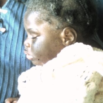
Chediak H Blood Smear
Please click to enlarge.
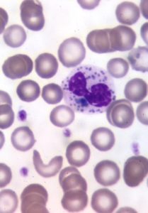
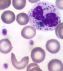
Note what signs you see.
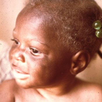
I (W. Wertelecki, M.D.) see a peripheral blood smear of a child with oculocutaneous partial albinism. The neutrophiles contain azurophilic and eosinophilic granules. In the image on the right and below is a cell shaped like a sickle (the patient has African ancestry). Please, see several additional illustrations of this blood smear.
PERSPECTIVE: In the context of clinical presentation, see related images where the diagnosis of C-H syndrome is virtually certain.
