
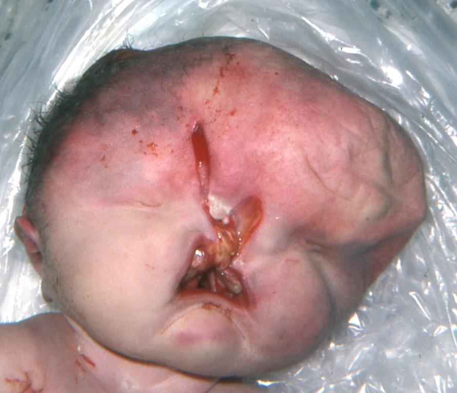
Encephalocele – Holoprosencephaly?
Please click to enlarge.
Note what signs you see.
I (W. Wertelecki, M.D.) see a classic example observed by Professor Peter Dignan of an infant illustrated the impacts of an altered development of the PROSENCEPHALON (forebrain). The posterior area of this brain vesicle devides or separates into the diencephalon (thalamus, hypothalamus, subthalamus, epithalamus and pre-tectum) and the forward area becomes the telencephalon which will evolve into whole CEREBRUM CORTEX, white matter and basal ganglia. Between the 5th – 8th weeks the forebrain devides into the left and right cerebral hemispheres. When such does not happen, the result is HOLOPROSENCEPHALY (HOL).
HOL results in a constallation of multiple malformations, some of which are illustrated here and others in a HOL image gallery and other pages (please search for the full keyword “holoprosencephaly”).
I see in this illustration a frontral protrusion that may be an “encephalocele”: the right palpebral fissure appears narrow, the occular globe small, and the orbit position hyperteloric (to far from the midline). The rudimentary nose is composed of separate (unfused) right and left anlage; the right anlage is not fused with the orbit; on the right there is an anlage of the nostril and its opening; the palate is cleft – the above interpretations are hypothetical. In any case, this image illustrates the correlation of the development of the prosencephalon with that of the facial and oral midline.